Notes For All Chapters Biology Class 11 CBSE
Each and every organism begins its life from single cell and grows to form large organism. All living cells grow in size and reproduce by dividing into two daughter cells, i.e., each parental cell divide and give rise to two daughter cells each time it divide, which again grow, divide and give rise to new cell population.
Therefore, such cycles of growth and division allows a single cell to form a structure that consists of millions of cells. Hence, the development of a multicellular organism from the unicellular zygote is achieved by the process of cell division.
Topic 1 Cell Cycle and Mitosis
Cell Cycle
Cell division is an essential process in all living organisms. The mode of cell division is fundamentally similar in all organisms. During the process of division of a cell, the processes like DNA replication and cell growth must take place in a sequential and coordinated manner to ensure the correct division and formation of progeny cells with intact genomes.
Cell cycle is an orderly sequence of events or a set of stages by which a cell duplicates its genome, synthesises the other constituents (important for the cell) of the cell and eventually divides into two daughter cells.
In terms of cytoplasmic increase, cell growth is a continuous process, while DNA synthesis occurs during a specific stage of the cell cycle only. The DNA (chromosomes) are further distributed to daughter nuclei by a series of events. All these events are well coordinated and are under __ genetic control.
Phases of Cell Cycle
A typical eukaryotic cell (human cell) divides once in every 24 hr. This duration of cell cycle can vary from an organism to organism and also from one cell type to another, e.g., Yeast cell progress through the cell cycle in only about 90 min.
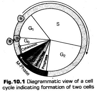
The cell cycle is divided into two phases
i. Interphase
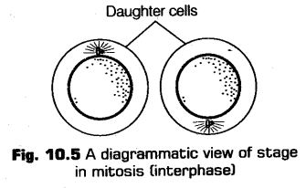
It is the period between the end of one cell division to the beginning of the next cell division, i.e., (between two successive M-phase). As no visible changes are observed at this stage, interphase is said to be Besting stage.
Daring this phase, cell prepares itself for both cell growth and DNA replication in an orderly manner. So, it is also known as preparation phase. It lasts for about 90-§6%, i.e., more than 95% of the total duration of cell cycle.
Interphase is further divided into fallowing three sub-stages on the basis of various synthetic activities
a. G1(Gap-l)-phase
It corresponds to the duration between the mitosis (M-phase) and initiation of replication of DNA. The cell become metabolically active during this period, grows continuously and prepares itself for DNA replication but does not undergo DNA replication.
b. S (Synthesis)-phase
It is known to be the phase in which actual synthesis or replication of DNA takes place. The overall amount of DNA doubles per cell, but no increase in chromosome number is seen during this stage, e.g., If the initial amount of DNA is 2C it will become 4C.
In case of animal cell, during S-phase DNA replication begins inside the nucleus while, the duplication of centrioles takes place in the cytoplasm.
c. G2(Gap-2)-phase
This phase is also called post-synthetic or pre-mitotic phase. During this stage the synthesis of DNA stops and proteins required for mitosis are being synthesised while, the growth of cell continues. It prepares the cell to undergo division.
Repair of damaged DNA and duplication of mitochondria, centrioles and plastids takes place during this phase.
G0-phase (Quiescent Stage)
This is known as the stage of inactivation of cell cycle. Cells in G0-phase remains metabolically active but do not proliferate, i.e., do not grow or differentiate till the time they do not get instruction to do so depending upon the requirement of an organism.
In adult animals, some cells like heart cells or nerve cells do not undergo division and many other cells divide only occasionally, to replace cells that have already _ been lost (because of some injury or cell death).
After completing G2-phase a cell may enter either into the G0-phase or into the M-phase directly. The duration of G0-phase may vary from indefinitely long to very short, except the nerve, bone and heart cells of chordates that are in permanent G0-phase.
a. M-phase
Following the interphase, the cell enters the M-phase or mitotic phase.
The M-phase may include any of the type of cell division i.e., mitosis or equational division and meiosis or reductional division. However, in general M-phase refers to mitotic phase.
M-phase also orderly involves the distribution of cell organelles and various macrobiomolecules to the daughter cells.
Mitosis
In this type of division the chromosomes replicate themselves and gets equally distributed into daughter nuclei, i.e., the chromosome number in the parental and progeny cell (diploid) become the same. Therefore, it is also known as equational division. Mitosis is also known as somatic cell division because it results in the formation of somatic cells.
Mitotic cell division is seen in the diploid somatic cells in animals, whereas, in plants; mitotic divisions is seen in both haploid and diploid cells.
Mitosis is considered to be the short period of chromosome condensation, segregation (karyokinesis) and cytoplasmic division.
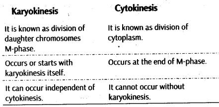
It is known to be the phase of actual cell division, which starts with the division of nucleus, followed by the separation of daughter chromosomes, i.e„ karyokinesis and terminates with the cytoplasmic division,
i. e., cytokinesis.
Karyokinesis
It is further divided into four main sub-stages, i.e., prophase, metaphase, anaphase and telophase.
i. Prophase
It is known to be the longest and the most complex phase of cell division because it lasts for about 50 min of total duration of mitotic phase.
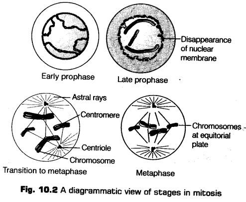
This is the first stage of mitosis that follows by the S and G2-phase of interphase. This phase is known for the initiation of condensation of chromosomal material, which during the process of chromatin condensation becomes untangled, and finally the centriole (already duplicated during S-phase of interphase) begins to move towards the opposite poles of the cell.
For the suitability in study we can categoriesprophase as
a. Early Prophase
* During this phase, condensation of chromosomal material takes place in order to form a compact mitotic chromosomes composed of two chromatids which are attached together at centromere.
* The most conspicuous change that take place during prophase is the formation of mitotic spindle. The initiation of mitotic spindle assembly, the micro¬tubules and the proteinaceous components of the cell cytoplasm helps in the completion of the process.
The mitotic spindle is formed between the two pairs of centrioles that migrate towards the opposite poles of the cell.
b. Late Prophase
At the end of the prophase, i.e., during late prophase the nucleolus disintegrates gradually and the nuclear envelope disappear. This disappearance marks the end of the prophase.
If we view cells under the microscope, during the prophase the cell will not show nucleolus, nuclear envelope, Golgi complex, endoplasmic reticulum, etc.
ii. Metaphase
It is the phase that starts after the disintegration of nuclear envelope in the late prophase. The chromosomes spreadout through the cytoplasm of the cell and are seem to be slighdy shortest and thickest of all. The chromosome can be easily observed under the microscope.
Mitotic Poison: Colchicine
Colchicine, an alkaloid is extracted from the corms of autumn crocus (Colchicum autumnale), acts as a poison for mitosis as it does not allow the formation of mitotic spindle to takes place by preventing the assembly of microtubules. But does hot affect replication of chromosomes. Thus, meristematic cells treated with this chemical show doubling of chromosomes. It usually causes arrest at metaphase of mitosis.
Each chromosome at this stage is made up of two longitudinal threads (sister chromatids) and are held together by the centromere in the centre. At the surface of each centromere disc-shaped structures called kinetochores are present, which helps in the attachment of spindle fibres to the chromosomes.
The chromosomes finally arrange themselves at the equator in one equitorial plane known as metaphase plate.
Following changes are observed during metaphase
(a) Attachment of spindle fibres to kinetochores of chromosomes.
(b) Movement of chromosomes to spindle equator and its
alignment along the metaphase plate through spindle fibres to both poles.
Note:
Kinetochore are the small disc-shaped structures at the surface of the centromeres, which serve as the sites of attachment of spindle fibres to the chromosomes that are moved into position at the centre of the cell.
Chromosomes are attached to the polar fibres at the kirtetochores, through kinetochore fibres.
iii. Anaphase
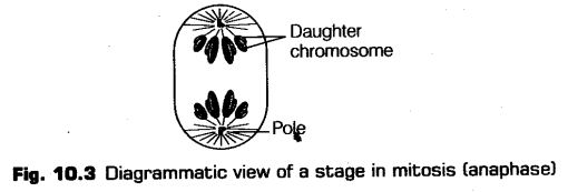
It is known to be the shortest duration phase, i.e., only of 2-3 min and is also very simple stage. At the beginning of this phase, splitting of chromosomes (that are already arranged at metaphase plate) takes place.
The two daughter chromatids now becomes the chromosomes of future daughter nuclei and start migrating towards the opposite poles along the path of their chromosome fibres.
Thus, following changes are observed during anaphase
(a) Splitting of centromeres and separation of chromatids.
(b) Movement of chromatids towards the opposite poles.
iv. Telophase
This is considered to be long and complex phase like prophase the final stage of mitosis. At the onset of this stage, the spindle disappears (absorbed in cytoplasm) and the chromosomes decondense and further loses their individuality after reaching their respective poles. In general terms, the events of prophase occurs just in reverse sequence during this phase.
Now the, individual chromosomes can not be seen and the chromatin material gets collected in the form of mass in both opposite poles.
Thus, following changes are observed during telophase
(a) The chromosomes gradually uncoil and cluster at opposite spindle poles. Thus, their individual identity as discrete elements is lost.
(b) Nuclear envelope slowly reformed around group of chromosomes.
(c) Reappearance of nucleolus, Golgi complex and ER also takes place.
Cytokinesis
It is commonly known as the division of cytoplasm of the parent cell into two daughter cells after the division of nucleus or karyokinesis. It occurs, so that the each daughter cell can receive nucleus of its own.
Thus, mitosis is not only the segregation of nucleus into two daughter nuclei, infact, the cell also divide itself into two daughter cells.
Differences between Karyokinesis and Cytokinesis
Cytokinesis in Animal Cells
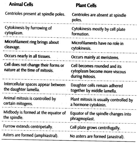
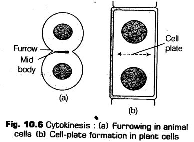
In animal cells, cytokinesis starts at metaphase. They typically divide by furrowing or by the appearance of furrow in the plasma membrane. This is also known as cleavage.
Due to the contraction and development of micro- filaments, a constriction develops which further deepens in a centripetal way known as cell furrow.
The furrow starts deepening gradually during telophase and finally gets joined in the centre by dividing the cytoplasm into two.
Cytokinesis in Plant Cells
Cytokinesis in plant cells is different from that in the animal cells due to the presence of a solid, rigid and in- extensible cell wall on the outside of the cell, the plant cell cannot undergo cytokinesis by the furrowing method. Therefore, plant cell divides by the cell-plate method. The formation of cell-plate usually begins during the late anaphase or early telophase. The formation of a new wall in plant cells takes place in the centre of the cell and start growing outward towards the opposite sides in order to reach the already existed lateral walls.
This new cell wall with the formation of simple precursor grows until it reaches the actual cell walls.
Once cell plate has divided the cell into two cells, it will continue to grow and develop into a new cell organelles (like mitochondria and plastids) and also gets distributed among two daughter cells during the cytoplasmic division or cytokinesis.
Significance of Mitosis
Mitosis has following significance
(i) It helps in the production of diploid daughter cells with equal and identical genetic complement.
(ii) Mitosis helps in growth of multicellular organisms.
(iii) It also helps in maintaining a proper cell size by dividing an overgrown somatic cell.
(iv) It is helpful in cell repair mechanism, eg., continuous replacement of the cells like of the upper layer of the epidermis, cells of the lining of the gut, and blood cells.
(v) The growth of multicellular organism is due to mitosis as, it is known that a cell cannot grow in size beyond a certain limit without disturbing the ratio between the nucleus and the cytoplasm.
After reaching the particular size, the cell divides in order to restore the nucleocytoplasmic ratio. Therefore, the growth of cell takes place by the increase in the number of cells, rather than the increase in the size of the cell.
(vi) It is also helpful in producing new cells for healing wounds and for regeneration.
(vii) Mitosis plays an important role in continuous growth of plants throughout their life by meristematic tissues like the apical and the lateral cambium.
Differences between Mitosis in Animal and Plant Cells
Meiosis is the phenomenon which occurs in any life cycle that involves the process of sexual reproduction. The production of offspring by sexual reproduction involves the fusion of two gametes (each having a complete haploid set of chromosomes).
Thus, meiosis is known to be the specialised form of cell division which reduces the chromosome number in such a way that each daughter nuclei receive only one set of each kind of chromosome, {i.e., maternal and paternal). It results in the production of haploid daughter cells. In meiosis, the nucleus divides twice but the replication chromosome takes place only once.
Thus, it is also known as the reductional division. In case of diploid organisms, meiosis takes place during the formation of spores or gametes whereas, in haploid organisms it takes place during germination of zygote.
Meiosis ensures the production of haploid phase in the life cycle of sexually reproducing organism whereas, the fertilisation restores diploid phase.
During this division, the homologous chromosomes of each pair separates from each other and reaches separate daughter cells which thereby reduces the number of chromosomes from diploid to haploid, i.e., from 2n to n. Thus, it is known as heterotypic division. It is farther divided into four phases,
i. e., prophase-I, metaphase-I, anaphase-I and telophase-I.
Prophase-I
It is considered to be the most complicated and prolonged phase as compared to the similar stage in mitosis.
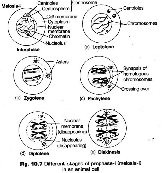
This phase is further sub-divided into five sub-phases on the basis of chromosomal behaviour, i.e., leptotene, zygotene, pachytene, diplotene and diakinesis.
i. Leptotene
It is known to be the very first stage of meiotic division following the interphase.
Following features are seen during this phase
* Chromosomes becomes gradually visible under light microscope.
* Centrioles start moving towards opposite ends or poles and ’ each centriole develops astral rays.
* Each chromosome is attached to the nuclear envelope through the attachment plate at both of its ends.
The essential features of meiosis are as follows
(а) Meiosis undergoes two successive cycles of nuclear and cytoplasm division, i.e., meiosis-I and II, but no DNA replication will result prior to second meiotic division.
(b) Meiosis-I and II occurs one after the another with a very short or no interphase.
(c) After the replication of parental chromosomes, initiation of meiosis-I takes place in order to produce identical sister chromatids at S-phase.
(d) Meiosis takes about days to gets completed instead of hours or minutes that are needed for mitosis.
(e) Pairing of homologous chromosomes and recombination takes place during meiosis.
(f) Finally, after the second meiotic division four haploid cells are being formed.
Homologous Chromosomes
There are two sets of chromosomes in a diploid cell undergoing meiosis, one set contributed by the male parent and the other by the female parent. There are always two similar chromosomes, having the same size, shape and position of centromere. In some organisms, the chromosomes give beaded appearance due to the presence of chromomeres (swollen area).
ii. Zygotene
This is the next sub-stage that takes place after the completion of the previous one. This is also a short lived stage like leptotene.
Following changes are seen during this phase
* Homologous chromosomes pair up. This pairing is done in a such a way that the genes of the same character present on the two chromosomes lie exactly opposite to each other. This process of association is known as synapsis.
* It is revealed from the electron micrographic studies that the formation of synaptonemal complex takes place by a pair of homologous chromosomes that show synapsis. The complex so formed, on account of synapsis forms a bivalent or a tetrad.
* The number of bivalents are half the total number of chromosomes and are not clearly visible at this stage.
iii. Pachytene
This is the stage which immediately follows zygotene where the pair of chromosomes become twisted spirally around each other and cannot be distinguished separately. This stage is comparatively long lived as compared to the previous two stages.
Following changes are seen during this stage
* Bivalent chromosomes clearly seen as tetrads.
* In this stage, sometimes exchange of genes or crossing over between the two non-sister chromatids of homologous chromosomes occurs at the points called recombination nodules, which appear at intervals, on synaptonemal complex. By the end of pachytene recombination gets completed leaving the chromosomes linked at the sites of crossing over.
Crossing Over
In this process, exchange of genetic material takes place between the non-sister chromatids of two homologous chromosomes. It finally leads to recombination of genetic material on the two chromosomes.
iv. Diplotene
It is the stage of longest duration of all. Following changes are observed during this stage
* In this the synaptonemal complex appears to get dissolved while, the chromatids of each tetrad remain clearly visible.
* Recombined homologous chromosomes of the bivalents get separated and form chiasmata (X-shaped structures).
* Chiasmata formation is necessary for the separation of homologous, chromosome which have undergone the process of crossing-over.
Diplotene can last for months or years in oocytes of some vertebrates.
v. Diakinesis
This is known to be the final stage of meiotic prophase-I. Also known as terminalisation, due to the shifting of chiasmata towards the end of the chromosomes. Following changes are observed during this stage
* Chromosomes become fully condensed.
* Nucleolus degenerates.
* Breakdown of nuclear envelope into vesicles.
* Formation of meiotic spindle (as in mitosis) in order to prepare the homologous chromosomes for separation.
* Diakinesis is the phase which represents the transition from prophase to metaphase of meiosis-l.
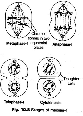
Metaphase-I
It is the stage followed by prophase (same as mitosis). Following changes are observed during this stage
(a) The bivalents during this phase arrange themselves on the two parallel equatorial plates.
(b) The centromeres project litde bit towards periphery. Since, there are two centromeres in each bivalent. Thus, each centromere is joined by chromosomal fibres.
(c) The fibres of the homologous chromosomes are always in the opposite directions.
Anaphase-I
This is the next phase after metaphase-I in which homologous chromosomes break their connection with each other and get separated.
This process of separation of homologous chromosomes is known as disjunction. The separated chromosomes are univalents and are also called dyads.
On reaching at the end of the anaphase, the two groups of chromosomes are produced (with each having half number of chromosomes).
The sister chromatids remains attached at their centromeres on the separation of the homologous chromosomes .
Telophase-I
This is the last stage of meiosis-I in which the chromatids at each pole of the spindle usually remain uncoiled and get elongated.
Following changes are seen during this stage
(i) Homologous chromosomes reach at their respective poles!
(ii) Reappearance of nuclear membrane and nucleolus takes place.
Cytokinesis
It is die stage during which the cytoplasm and other organelles divides into two equal halves of cells.
Interkinesis
This is the stage betweenJbe two meiotic divisions, i.e., the meiosis-I and II. It is generally short lived. During this process, no replication of DNA occurs. It is necessary for bringing true haploidy DNA in daughter cells.
It is in fact considered as incipient interphase.
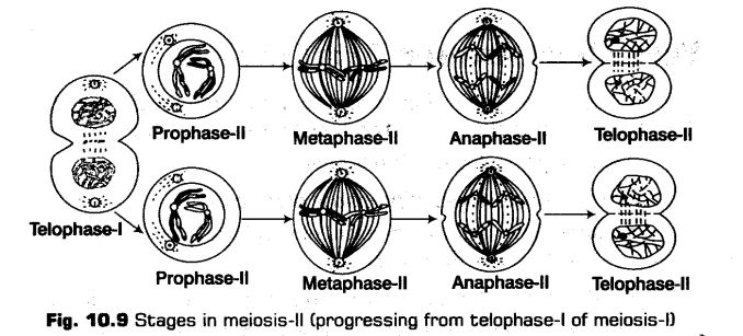
Meiosis-II
Meiosis-II is known by another term, i.e., homotypic division, because in this division chromosome number remains same, as produced in meiosis-I. It is initiated immediately after cytokinesis. It is often known an equational division. Meiosis-lf also resembles a normal mitotic division in contrast to meiosis-I because it distributes chromatids to daughter cells (like mitosis).
Prophase-ll
This is known to be the very short stage out of the all. During this process the chromosomes again become compact in organisation.
Following changes are seen during this process
(i) The centrioles duplicate themselves by the separation of the two members of the pair.
(ii) Each chromosome comprising two chromatids become visible in the nucleus. These chromosomes further become thick and short in size.
(iii) Nuclear envelope breaks down and the formation of spindle apparatus takes place and nucleoli disappears.
Metaphase-ll
This is the next phase followed immediately after prophase-II.
Follotving changes are seen during this process
(i) The chromosomes align at the equator or the metaphase plate, in the similar way as in_mitosis.
(ii) Chromosomes get attached to the fully formed spindle apparatus and the kinetochores of sister chromatids for each chromosome face the opposite poles and each is attached to the kinetochore microtubule coming from the pole of that side.
Anaphase-ll
This is the phase followed immediately after metaphase-II.
Following changes are seen during this process
(i) The centromere of each chromosome splits, that was holding the sister chromatids together.
(ii) The shortening of chromosomal microtubules take place, the two chromatids of each chromosome start moving away from each other and finally reaches the opposite poles of the spindle (now called chromosomes).
Telophase-ll
This is known to be the last stage of second meiotic division and show changes equally opposite to that of the prophase-II. Meiosis ends up with the progress of this particular phase, i.e., telophase-II.
Following changes are seen during this process
(i) Formation of a nuclear envelope (from ER) around each set of chromosomes.
(ii) Nucleoli reappears due to the synthesis of ribosomal RNA (rRNA) and ribosomal DNA (rDNA).
Cytokinesis
This occurs after every nuclear division. It includes separation of cytoplasm and organelles in the two halves of the cell. Thus, the two daughter cells formed have half the number of chromosomes and the amount of nuclear DNA. Both the cells undergo divisions and give rise to four cells. These haploid cells are arranged tetrahedrally and are collectively called tetrahedral tetrad.
It usually send equal amounts to each daughter cells but sometimes division is highly unequal (especially in egg production).
Note:
Cytokinesis may or may not occur between meiotic divisions, however, in most of the organisms it is of rare occurrence after Mitosis and Meiosis-ll.
Significance of Meiosis-ll
(i) It maintains the same chromosome number in the sexually reproducing organisms. From a diploid cell, haploid gametes are produced which in turn fuse to form a diploid cell. Haploid gametes are formed due to reduction of chromosomes to its half.
(ii) It restricts the multiplication of chromosome number and maintains the stability of the species.
(iii) Maternal and paternal genes get exchanged during crossing over. It results in variations among the offspring.
(iv) All the four chromatids of a homologous pair of chromosomes segregate and go over separately to four different daughter cells. This leads to variation in the daughter cells genetically.
(v) Paternal and maternal chromosomes assort independendy. Thus, cause reshuffling of chromosomes and traits controlled by them.
Differences between Mitosis and Meiosis

Leave a Reply