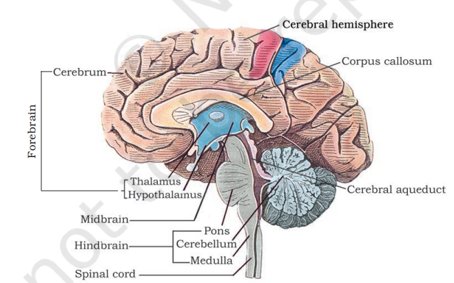
Q1. Briefly describe the structure of the brain.
Answer :
The brain is the central information-processing organ of the body, protected by the skull and cranial meninges (dura mater, arachnoid, and pia mater). It has three major parts:
1. Forebrain
The forebrain is the largest and most complex part of the brain, consisting of the following:
Cerebrum:
- Structure: Divided into two hemispheres (left and right) connected by a bundle of nerve fibres called the corpus callosum.
- Cerebral Cortex: The outer layer of the cerebrum, also called grey matter, has folds to increase surface area. The inner layer is made of white matter, which contains myelinated nerve fibres.
- Functions: Responsible for sensory perception, voluntary motor actions, reasoning, memory, and emotions.
Thalamus:
- Structure: Located beneath the cerebrum.
- Function: Acts as a relay centre, processing and transmitting sensory and motor signals to the appropriate areas of the cerebrum.
Hypothalamus:
- Structure: Located at the base of the thalamus.
- Functions:
- Regulates body temperature, hunger, thirst, and circadian rhythms.
- Contains neurosecretory cells that produce hypothalamic hormones.
- Plays a role in emotional responses and motivation.
2. Midbrain
The midbrain is a small region that connects the forebrain and hindbrain.
Structure: Contains a canal called the cerebral aqueduct and four rounded structures known as the corpora quadrigemina.
Functions:
- Integrates sensory inputs (visual, auditory, and tactile).
- Coordinates reflex movements of the head, neck, and eyes.
3. Hindbrain
The hindbrain comprises the following structures:
Cerebellum:
- Structure: Has a highly folded surface to accommodate more neurons.
- Functions: Maintains balance, posture, and coordination of voluntary movements.
Pons:
- Structure: Located between the midbrain and medulla.
- Functions: Connects various parts of the brain and helps in controlling respiration.
Medulla Oblongata:
- Structure: Connects the brain to the spinal cord.
- Functions:
- Controls vital reflexes such as heartbeat, breathing, and digestion.
- Regulates involuntary actions like swallowing and sneezing.
- Functions:
Q2. Compare the following:
Answer :
(a) Central Neural System (CNS) vs. Peripheral Neural System (PNS):
| Feature | CNS | PNS |
|---|---|---|
| Definition | Includes the brain and spinal cord, acting as the main control centre. | Includes all nerves outside the CNS, connecting it to the rest of the body. |
| Components | Brain and spinal cord. | Cranial nerves, spinal nerves, and ganglia. |
| Function | Processes and interprets information. | Transmits signals between the CNS and body parts. |
| Divisions | Not divided further. | Divided into somatic and autonomic systems. |
(b) Resting potential and action potential
| Feature | Resting Potential | Action Potential |
|---|---|---|
| State | Neuron is at rest, polarised. | Neuron is active, depolarised. |
| Charge | Inside of axon is negative, outside is positive. | Inside becomes positive, outside negative. |
| Ion Movement | inside, outside due to sodium-potassium pump. | Rapid influx of , followed by efflux. |
| Function | Maintains neuron readiness. | Transmits nerve impulse. |
Q3. Explain the following processes:
(a) Polarisation of the membrane of a nerve fibre
(b) Depolarisation of the membrane of a nerve fibre
(c) Conduction of a nerve impulse along a nerve fibre
Answer :
(a) Polarisation:
- At rest, the axon membrane is more permeable to
and impermeable to
.
- Sodium-potassium pumps actively transport
out and
in, maintaining a negative charge inside.
- This results in a resting membrane potential of approximately
.
(b) Depolarisation:
- When a stimulus reaches the membrane,
channels open, and
rushes inside.
- This reverses the polarity of the membrane (inside becomes positive, outside negative).
- This phase is called depolarisation, and it generates an action potential.
(c) Transmission across a chemical synapse:
- Action potential arrives at the presynaptic terminal, triggering
channels to open.
influx causes synaptic vesicles to release neurotransmitters into the synaptic cleft.
- Neurotransmitters bind to receptors on the postsynaptic membrane, opening ion channels.
- Depending on the neurotransmitter, the postsynaptic neuron is either excited (action potential generated) or inhibited.
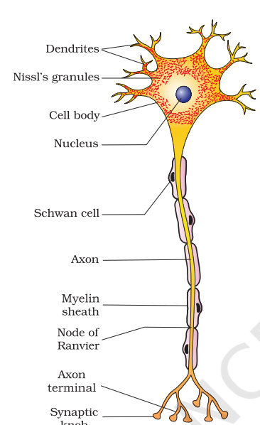
Q4. Draw labelled diagrams:
(a) Neuron
(b) Brain
Q5. Write short notes:
Answer :
(a) Neural Coordination:
- Neural coordination involves the nervous system regulating and integrating body functions through electrical signals.
- The CNS and PNS work together to maintain homeostasis and respond to internal and external stimuli.
(b) Forebrain:
- Composed of the cerebrum, thalamus, and hypothalamus.
- Controls sensory perception, voluntary actions, emotions, hunger, and body temperature.
- The cerebral cortex processes higher cognitive functions like learning and memory.
(c) Midbrain:
- Located between the forebrain and hindbrain.
- Contains the cerebral aqueduct and corpora quadrigemina.
- Integrates sensory inputs and coordinates reflex movements.
(d) Hindbrain:
- Includes the cerebellum, pons, and medulla oblongata.
- Controls balance, posture, respiration, and reflex actions like sneezing.
(e) Synapse:
- A junction where a nerve impulse is transmitted from one neuron to another.
- Includes electrical and chemical synapses, with neurotransmitters playing a key role in the latter.
Q6. Mechanism of Synaptic Transmission
Answer :
- Presynaptic Events:
- Action potential arrives at the axon terminal.
- Voltage-gated
channels open, causing an influx of
.
- Synaptic vesicles release neurotransmitters into the synaptic cleft.
- Synaptic Cleft:
- Neurotransmitters diffuse across the cleft to the postsynaptic neuron.
- Postsynaptic Events:
- Neurotransmitters bind to receptors, opening ion channels.
- Excitatory neurotransmitters generate an action potential; inhibitory ones prevent it.
Q7. Role of
in the Generation of Action Potential
Answer :
- At rest,
is concentrated outside the axon.
- Upon stimulation,
channels open, allowing
to rush in.
- This depolarises the membrane, creating an action potential.
- The rapid influx of
is essential for nerve impulse transmission.
Q8. Differentiate Between :
(a) Myelinated vs. Non-myelinated Axons:
| Feature | Myelinated Axons | Non-myelinated Axons |
|---|---|---|
| Structure | Covered with myelin sheath. | Not covered with myelin sheath. |
| Conduction | Saltatory (faster). | Continuous (slower). |
| Location | Found in spinal and cranial nerves. | Found in autonomic nerves. |
(b) Dendrites vs. Axons:
| Feature | Dendrites | Axons |
|---|---|---|
| Function | Transmit impulses toward cell body. | Transmit impulses away from cell body. |
| Structure | Short and branched. | Long and unbranched. |
(c) Thalamus vs. Hypothalamus:
| Feature | Thalamus | Hypothalamus |
|---|---|---|
| Function | Relays sensory and motor signals. | Controls temperature, hunger, and emotions. |
| Location | Above the hypothalamus. | Below the thalamus. |
(d) Cerebrum vs. Cerebellum:
| Feature | Cerebrum | Cerebellum |
|---|---|---|
| Function | Higher cognitive functions. | Balance and coordination. |
| Structure | Largest part of the brain. | Folded surface for more neurons. |
Q9. Answer the Following
(a) Which part of the human brain is the most developed?
- The cerebrum is the most developed part of the brain.
- It handles complex processes like memory, learning, emotions, and voluntary movements.
(b) Which part of our central neural system acts as a master clock?
- The hypothalamus acts as the master clock.
- It regulates circadian rhythms, controlling sleep-wake cycles and hormone secretion.
10. Distinguish between :
(a) Afferent vs. Efferent Neurons:
| Feature | Afferent Neurons | Efferent Neurons |
|---|---|---|
| Function | Carry impulses to the CNS. | Carry impulses from the CNS. |
| Direction | Toward CNS. | Away from CNS. |
(b) Impulse Conduction in Myelinated vs. Non-myelinated Fibres:
| Feature | Myelinated Fibres | Non-myelinated Fibres |
|---|---|---|
| Speed | Faster due to saltatory conduction. | Slower due to continuous conduction. |
(c) Cranial vs. Spinal Nerves:
| Feature | Cranial Nerves | Spinal Nerves |
|---|---|---|
| Number | 12 pairs. | 31 pairs. |
| Function | Control head and neck functions. | Control body and limb functions. |


Leave a Reply